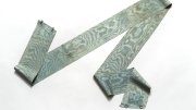

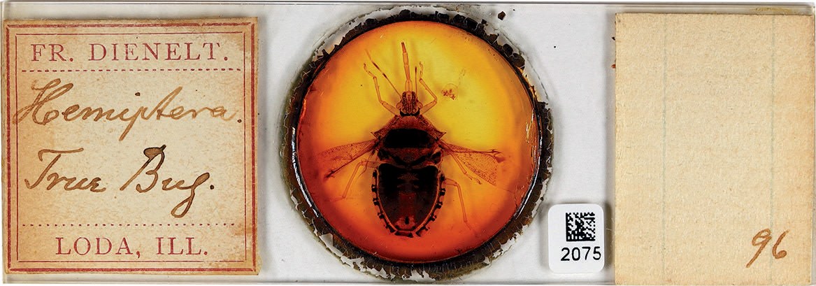
For the last two years, curators have scoured the dusty corners and cabinets of Harvard’s Museum of Comparative Zoology (MCZ), collecting about 50,000 microscope slides featuring tiny specimens. Some, like the translucent wings of a true bug (Heteroptera Latreille, hemiptera) and segments of a Western honeybee (Apis mellifera), are more than a century old.
Professor of organismic and evolutionary biology Mansi Srivastava, one of the MCZ’s curators of invertebrate zoology, especially likes a series of slides of squid embryos, collected in 1880 by marine biologist William Keith Brooks, Ph.D. 1875. “It’s the first time that the detailed anatomy of squid embryos is captured at different stages,” she says. “It’s just amazing—the fact that we have the actual objects, the actual tissue of those squid embryos that became the basis of our understanding of how nature makes a squid.”
Srivastava has led the continuing curation project to restore, catalog, and digitize the MCZ’s slides. The goal, she says, is to make this “meaningful data available to anyone anywhere on the planet.” Meanwhile, anyone can see a small selection of these historic treasures, artful in their own right, at the Harvard Museum of Natural History. Making the Invisible Visible: Digitizing Invertebrates on Microscope Slides highlights eight slides visitors can view through a microscope, among them a butterfly proboscis (through which the insect draws fluids) and a snail radula (a flexible ribbon of thousands of microscopic teeth by which the mollusk grates up food). Fifteen enlarged digital-print photographs on a nearby wall reveal the intricacy of a caterpillar’s skin, an ant’s head, and a veined dragonfly wing.
Historically, the slides also reflect how scientists communicated their findings. See the image of soft coral tissue on which pioneering zoologist Addison Emery Verrill, S.B. 1862, etched the words, “sent to James Dwight Dana by Charles Darwin.” Professor of entomology William Morton Wheeler (the “ant man” who predated Harvard’s late Pellegrino University Professor E.O. Wilson) illustrated embryonic forms of the praying mantis (Stagmomantis carolina) for his 1893 thesis—based on the use of a microscope. The revolutionary instrument, invented in the late 1500s, enabled scientists to see the minutest of creatures and describe the anatomy and morphology that helped explain whole new species. In this sense, says Srivastava, the MCZ’s slide collection evokes an age of discovery, offering an “incredible glimpse into things that you couldn’t have really imagined beforehand.”