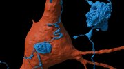
Might memories and habitual actions be hardwired into the brain’s physical structure? Knowles professor of molecular and cellular biology Jeff Lichtman thinks it’s likely. He and colleagues spent the past decade analyzing one cubic millimeter of cerebral cortex (about the volume of a poppy seed) to create the first detailed map of connections in the human brain. Their analysis has revealed numerous complex structures of great beauty and unknown purpose: nerve fibers growing in whorls, as if circling in place as they grew before settling on a path to connect with other cells; a new class of neurons with receptors (dendrites) that point only in two, diametrically opposed directions, and aligned with nerve fibers in an unconnected, underlying layer of white matter (none of the scientists has even a theory as to why this alignment exists); and powerful multisynaptic connections, rare instances when nerve fibers make as many as 50 or more connections to a single cell.
Their observations have upended assumptions about how the brain is wired. “Many people expect science to resolve, to answer, to cure,” reflects Lichtman, but in this case, simply describing nature has revealed that the brain is “more complicated than we thought.”
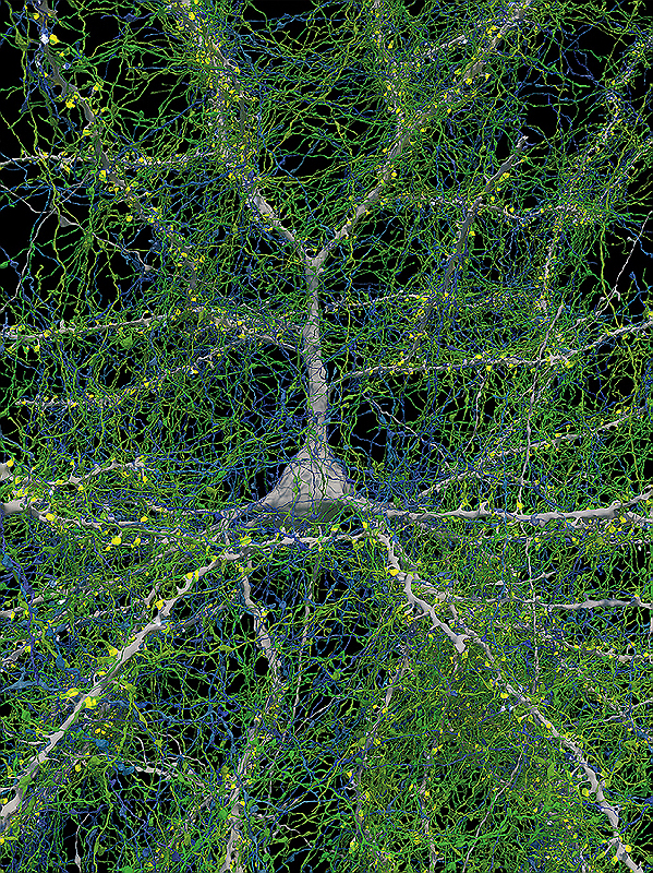
They discovered that glial cells, which protect nerve cells, outnumber neurons two to one. But much of what the researchers found eludes explanation. Most of the connections in the brain can be characterized as weak—a single synaptic connection from a nerve fiber to a cell. But Lichtman and colleagues observed a few very strong connections with many synapses for every cell, that would allow information to flow through the brain very quickly. “And maybe,” says Lichtman, “that is what learned information is, what memories actually are.”
Techniques in Brain Mapping
Surgeons removed the tissue used in the analysis from the temporal lobe of a 45-year-old woman to gain access to an underlying lesion. After infusing and preserving the sample with a hardening resin, the researchers used a diamond knife to slice it into more than 5,000 sections, thinner than a thousandth of a human hair. “Any thicker,” says Lichtman, “and we can’t actually follow the wires through.” Each slice was scanned with a purpose-built multibeam electron microscope (the world’s first when the experiment began) at a resolution so high, he said, that to see all the detail in just one slice would require an array 750 laptop screens wide by 500 high. Creating the images took six months, running the equipment 24 hours a day, seven days a week. Then came the hard part.
To generate the 1.4 petabyte dataset describing the wiring of this millionth-part of a human brain, researchers at Google, led by neuroscientist Viren Jain, developed novel neural networking techniques to detect the objects within each slice and stitch them together to render a three-dimensional space. They used “flood-filling neural network” technology to color the cells, nerve fibers, and blood vessels, Lichtman explains, “basically pouring paint into these objects across many sections.” The seed-sized sample they imaged contains 57,000 cells, 230 millimeters of blood vessels, and 150 million synapses, the junctions between neurons where electrical and chemical signals are transmitted. Lichtman says in idle moments, instead of playing solitaire, he navigates the publicly available inner space they’ve mapped using a tool called Neuroglancer.
With faster equipment and a greatly expanded group of global collaborators, Lichtman next wants to map the entire brain of a mouse, which would generate a thousand times more data. Mapping the whole human brain is not yet possible: the data produced would overwhelm current storage systems. But a basic question that such work would help answer is how the human brain differs from those of other animals.
Theories on Memory Storage and Learning
Lichtman suspects that the main difference is the abundance of associational cortex in humans, the tissue primarily devoted to storing information. “To put it another way,” he says, “humans are the adult animal with the least amount of circuitry that is entirely designed based on genes.” Instead, human brain circuitry is shaped by what has been learned. “Humans are obligate learners,” he continues. A human raised without sight, who is then permitted to see, for example, can look at a triangle and perceive the image, without understanding what it is. “They have to place their finger at one apex, and then count how many times they move their hand to know it is a triangle. They don’t gestalt it as a triangle. It’s like trying to understand the meaning of a word when you are just starting to read, and you have to phonetically sound out each letter to know what the word is. That’s something not evident in other animals, this enormous requirement for learning in order to do even the basic tasks of thinking, because we think in a language that is learned.”
Lichtman, recently named dean of science in the Faculty of Arts and Sciences, believes that this learning is embedded in the physical structure of the brain’s wiring. “I don’t see what the alternative would be,” he says. “If brain activity stops, as when a child suffers anoxia after falling into a frozen pond,” but rescuers get the heart beating again, “all the memories already stored are stably maintained.” A once-popular theory, that memories are stored as protein molecules, hasn’t withstood scrutiny. On the other hand, the superabundant connections in the brain, he said, “can certainly store information.” Lichtman theorizes that the rare neurons with many synaptic connections he and his colleagues observed during their mapping project represent things that have been learned, such as reflexively “taking your foot off the gas and putting it on the brake automatically” at a red light.
Observational science of the type that these researchers are pursuing is sometimes characterized pejoratively. But “Darwin spent a huge amount of time looking at finch beaks and shell shapes—that’s why he came up with a really good hypothesis,” Lichtman says. Sometimes, the best humans can do when confronting the complexity of the natural world, he continues, is to describe it—until it is no longer surprising. In the case of their own research, he notes, “We have just made our first baby steps into these brains. And everything is surprising.”
