Museum specimens yield new secrets to non-invasive imaging technology

New technology lets researchers see differences inside museum specimens

An earthworm rendered in 3D, with transparent outer skin to show the internal structures

An earthworm rendered in 3D, with transparent outer skin to show the internal structures | Image courtesy of Rosa M. Fernández García and Alexander Ziegler

A still from a video shows the internal  the organs of an earthworm, including its four hearts, in blue.

A still from a video shows the internal the organs of an earthworm, including its four hearts, in blue. | Image courtesy of Rosa M. Fernández García and Alexander Ziegler

Colored chambers of the heart in a preserved larval axolotl, an endangered salamander

Colored chambers of the heart in a preserved larval axolotl, an endangered salamander | Image courtesy of Zachary Lewis

The skull of an anole from Hispaniola, showing the many bones in a lizard’s skull, as compared to a mammal’s

The skull of an anole from Hispaniola, showing the many bones in a lizard’s skull, as compared to a mammal’s | Image courtesy of Stephanie Pierce and Blake Dickson

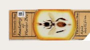
Even to a trained zoologist, one earthworm looks very much like the next. So much so, in fact, that one species generally can’t be distinguished from another just by looking—unless the animal has been painstakingly dissected. Exposing its internal morphology in this way destroys the specimen. The earthworm at right, with its life systems laid out in color, was “wild caught,” according to its record in Harvard’s Museum of Comparative Zoology (MCZ) specimen database, MCZbase. Aporrectodea caliginosa, a common species, was collected in the vicinity of 26 Oxford Street in Cambridge, where the museum is located. “You can imagine the drama” of its capture, quips James Hanken, Agassiz professor of zoology, curator in herpetology, and director of the MCZ.

What makes this lowly worm special is that it has been scanned using micro-computed tomography (microCT). The technology allows investigators to peer inside, to “see” the worm’s internal structures, including the digestive tract (pink), nervous system (yellow), and circulatory system (blue). Each can be studied separately and rotated in space in any direction to facilitate close inspection.

Before the MCZ tried it, no one had used a microCT scanner to image museum specimens, and the project has become an enormous success. The ability to see the reproductive organs, for example—the seminal vesicles, the number of testes and ovaries, and the spermathecae—has allowed researchers to dispel the “taxonomic chaos” that has plagued the study of earthworms.

But the applications are far broader. Such imaging allows the noninvasive study of extinct or critically endangered creatures in the MCZ’s 20-million-plus specimen collections, such as the larval axolotl at left above. Assistant professor in organismic and evolutionary biology Stephanie Pierce, curator of vertebrate paleontology at the MCZ, explains that one of her students has imaged the skulls of anole lizards (at left below) to look at how the inner-ear system (specifically, the delicate vestibular system so crucial for balance and spatial orientation, not depicted here) relates to the braincase bones, and how the vestibular system changes with habitat segregation on islands. For the MCZ, home to the largest university-based natural history collection in the world, treasured specimens can now be safely examined and shared with researchers and students, illustrators, artists, conservationists, and teachers worldwide.


Special thanks to the Radcliffe Institute for Advanced Study, which held a symposium, “University as Collector,” in which the earthworm scans were shown to the public, and especially to the Museum of Comparative Zoology.

Read more articles by Jonathan Shaw

You might also like

Tk tk Iran

Artist Azadeh Akhlaghi reconstructs moments of Iranian political upheaval in a series of meticulously staged images.

Science and art capture the microscopic natural world.

George Washington’s Sash on Display at Peabody Museum Starting May 25

A famous American fashion statement helps bring Revolutionary history to life.

Most popular

An animal’s journey from grief to love shows how much humans need each other, too.

Meet Harvard’s 2026 Student Commencement Speakers

Two undergraduates and a Ph.D. candidate will address the graduating class on May 28.

Harvard Weathers a Year of Turmoil

The federal government has launched unprecedented actions against the University. Here’s a guide.

Explore More From Current Issue

Star-filled night sky with the Milky Way arching over a rocky silhouette.

There’s a growing movement to curb light pollution. It starts on your front porch.

A chaotic scene in a messy room with people engaging in various activities, some cleaning.

Until the 1950s, professionals cleaned up after students in the dorms.

Harvey Mansfield seated in a bright yellow chair, surrounded by bookshelves and cozy decor.

The retired government professor has been a rare conservative voice on campus for decades.